Evidence Collection
Evidence Collection From Victim
Documentation
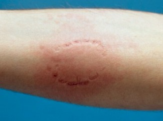
A record of the injury, including descriptive and narrative notes that document the physical appearance, colour, size and orientation of the injury. Questions such as ' What is the location on the body? What is the relative contour and elasticity of the site? Can the difference between marks from the upper and lower teeth be determined? What types of injuries are present? Cuts? Bruises? Scrapes?' are also answered through descriptive and narrative notes.
Photographs
Extensive orientation and close-up photographs are taken using a intra-oral camera. All pictures are taken in both colour and black-and-white. A reference scale, such as a ruler, is placed on the same plane as the injury and is visible in the photographs to show the measurement of the injury. The examiner has to make sure that the camera is directly over the injury.
Saliva Swabs
Saliva will have been left during biting and will have to be collected. A double swab technique is used to collect the sliva. First a cotton swab that is dampened with distilled water is wiped over the are that the bite occurred. Then the Analysts use a dry swab to collect any of the moisture left over. Both swabs are dried for at least fourty five minutes before they are allowed to be tested. A DNA sample is also taken from the victim and is compared to the saliva swabs.
Impressions
An accurate impression of the wound will then have to be made to show any irregularities made by the teeth. Analysts use vinyl polysiloxane, polyether or any other impression material that is used in a dental office. Dental Acrylic or plaster is also used as a rigid support for impression materials. It will allow the impression to accurately record the curvature of the skin.
Evidence Collection from Bite Suspect
Clinical Examination
An anylists will closely examine the intra-oral and the extra-oral structures of the bite suspects mouth. Any significant findings are noted. Results of specific examinations such as fractures, pocketing, etc. are documented by the analysts.
Photographs
Full facial and Profile pictures are taken in addition to intra-oral pictures to shoe the structures of the mouth. Pictures are also taken with a reference scale to help whith examination.
Impressions
It is necessary to produce extremely accurate impressions of the teeth to record all characteristics of the teeth. It is recommended that two sets of study molds be produced using a hard stone, such as dental die stone.
Bite Sample
A sample of the suspects bit is recorded in either a wafer of baseplate wax or in silicone putty material made for this use. The sample is photographed right after is is made for future reference to show that no change has occured. The suspects should be held until all of the samples and impressions are satisfactory.
Physical Comparison
The most common way to conclude that the suspect made the bitemark is to compare the patterns of the teeth to the impressions, bite samples, photographs, and notes taken in the clinical examination. The most accurate technique is a technique using the computer. Other comparison methods include direct comparison wiht the sus[ects study casts witht eh photographs of the bitemark, comparison of test bites made by the suspects teeth to the actual bitemark, and the use of radiographic imagingand scanning electron microscopy.
